Whereas unipolar neurons are generally confined to the ganglia of the brain and/or longitudinal nerve cords, cells of plexuses are dominated by bipolar cells.
From: Encyclopedia of Biodiversity, 2001
Smell and Taste
Andrea Giovanna Pineda, … Richard L. Doty, in Handbook of Clinical Neurology, 2019
Gross Anatomy
Although seemingly ubiquitous throughout the vertebrates, the NT is a multifaceted paired structure that varies among species in terms of morphology, chemistry, and neural connections (Von Bartheld, 2004). Peripherally it is commonly comprised of a group of thin unmyelinated bipolar and unipolar neurons embedded within autonomic and chemosensory nerve fascicles in the nasal cavity, as shown for the human infant in Fig. 9.3 (Wirsig-Wiechmann and Lepri, 1991; Wirsig-Wiechmann, 1993b). More centrally, it forms a plexus beneath the most rostral part of the brain, lying medial to the olfactory bulb and tract, as shown in Fig. 9.1 (Bojsen-Moller, 1975). In the rhesus monkey, the NT-related cluster of neurons enters the parenchyma of the brain along with the branches of the anterior cerebral artery (Silverman et al., 1982). Eisthen and Northcutt (1996) operationally defined this nerve as “the anterior cranial nerve that projects a loose group of fibers between the nasal region and the hypothalamic–preoptic region of the forebrain, passing through or over the surface of the olfactory bulb.” Aside from its projections to forebrain regions involved in reproductive function (Demski and Schwanzel-Fukuda, 1987), including the amygdala (Jennes, 1987), its central projections are largely unknown. In some species, such as the goldfish, they are widespread and involve the retina (Von Bartheld and Meyer, 1986).

Fig. 9.3. Diagrammatic sketch of the right side of the nervus terminalis and its extensions in an infant, seen from the median plane and projected onto the olfactory bulb and fila olfactoria. Depiction of ganglion cells is used to demonstrate where main aggregations are located. The branches of the NT are embellished in order to facilitate their identification.
From Brookover, C., 1917. The peripheral distribution of the nervus terminalis in an infant. J Comp Neurol 28, 349–360.
Despite being larger in humans than in most other species (Johnston, 1914), its characteristic thinness and close proximity to the olfactory bulb and tract makes the NT difficult to distinguish from these structures. Moreover, its relationship to trigeminal nerve endings within the nasal cavity is not clear, as its terminations are difficult to identify by electron microscopy. The NT is frequently extracted along with other parts of the brain during human autopsies and dissections, making it nearly impossible to subsequently locate (Bordoni and Zanier, 2013). Adding to this difficulty, the ganglion terminale, the central ganglion of the NT complex, is inconsistent in size and shape (Jennes, 1986). This ganglion has been suggested to be homologous in mammals to the nucleus olfactoretinalis found in fish (Demski and Schwanzel-Fukuda, 1987), a nucleus associated with interactions between the olfactory and visual systems. However, the general association between the NT and both the visual and olfactory systems is enigmatic in most forms. It is noteworthy that the NT is present in whales and dolphins—animals that lack olfactory bulbs and a functioning olfactory system (Buhl and Oelschlager, 1986).
In most forms, a defining element of the NT is its immunoreactivity to GnRH. Schwanzel-Fukuda and Silverman (1980) demonstrated the presence of GnRH within the NT of the guinea pig (Cavia porcellus). GnRH was absent from the olfactory and vomeronasal nerves, implying that the GnRH-secreting fibers are unique to the NT. In the rat, the NT likely supplies the main and accessory olfactory systems with GnRH fibers (Witkin and Silverman, 1983). Uptake studies in the mouse using horseradish peroxidase (HRP) suggest that the GnRH neurons of the NT have access to fenestrated capillaries within the subarachnoid space, the subepithelial connective tissue of the nasal mucosa, and the nasal epithelium proper (Jennes, 1986).
That being said, it should be emphasized that not all NT fibers are GnRH reactive (Schwanzel-Fukuda et al., 1986; Koza and Wirsig-Wiechmann, 2001). Moreover, GnRH does not appear to be present in the NT of some species, such as the lamprey (Northcutt and Puzdrowski, 1988), although a number of GnRH peptides, some of which are yet to be identified, may not be labeled by extant antibodies (Sherwood et al., 1993). Importantly, cell populations within the NT of various organisms have been shown to immunologically label a wide range of antibodies, including substance P (Kyle et al., 1995), choline acetyltransferase (Schwanzel-Fukuda et al., 1986), vasoactive intestinal polypeptide (Schwanzel-Fukuda et al., 1986), acetylcholinesterase (AChE) (White and Meredith, 1995), OMP (Valverde et al., 1993), calbindin (Abe et al., 1992), GABA (Fischer and Stell, 1997), tyrosine hydroxylase (White and Meredith, 1995), FMRF-amide (Wirsig-Wiechmann, 1990; Eisthen and Northcutt, 1996; Fiorentino et al., 2002), and neural cell adhesion molecule (NCAM) (Schwanzel-Fukuda and Pfaff, 2002). Nitric oxide synthase (NOS) coexists with AChE in the NT in some species, presumably related to a role in modulating the function of nitric oxide (Fuller and Burger, 1990).
The cells of the NT complex can be divided into two types: Type I, which are large cells immunoreactive to GnRH, and Type II, which are small cells not immunoreactive to GnRH (Oka and Ichikawa, 1991) (Fig. 9.2). In some chondrichthyes, as well as in lung fish, distinct divisions of the NT have been identified. In the lungfish, for example, an anterior division enters the brain via the olfactory bulb, whereas a posterior division enters into the diencephalon at the level of the optic nerve. While there is debate as to whether these divisions should be considered separate nerves (Schober et al., 1994), it appears likely that these primitive divisions were somehow incorporated into what is now considered the more unitary NT found in more advanced forms (Von Bartheld et al., 1988).
Read full chapter
URL:
https://www.sciencedirect.com/science/article/pii/B9780444638557000095
Cytology of the Central Nervous System
L. Jennes, in Conn’s Translational Neuroscience, 2017
Dendrites and Axons
Neurons have the unique property to receive, process, integrate, and transmit information over sometimes very long distances. Specific features associated with different parts of a neuron accommodate these tasks: dendrites usually receive a signal which is then propagated along the perikaryon to the axon which carries the signal to its terminal. In most neurons, many dendrites extend from different regions of the perikaryon and taper off with increasing distance from the cell body. Dendrites can branch extensively and develop dendritic trees, which form the portion of a neuron that receives most of the synaptic input. Often, dendrites contain small protrusions or spines along their surface, which cause a significant increase in the accessible surface area available for synaptic innervation (Fig. 1.3). The cytoplasmic composition of dendrites, especially at their base, resembles the one of the perikaryon and contains most organelles, such as the rER or lysosomes but usually not a Golgi apparatus. Dendrites contain many microtubules and neurofilaments as well as microfilaments, which are particularly dense in dendritic spines where the microfilaments are involved in the control of spine size and shape. The sER reaches sometimes the base of dendritic spines, which may also contain polyribosomes indicating that local protein synthesis can occur. When a neuron receives synaptic signals at a dendrite, these signals are integrated and propagated to the perikaryon in the form of an electrical signal, the synaptic potential. If this potential reaches a certain threshold, an all or a none response is initiated at the initial portion of an axon, the axon hillock, and an action potential is generated that travels unidirectionally along the axon to its terminal. Here, the arriving action potential causes the release of neurotransmitter from the presynaptic terminal. The axon hillock is a highly specialized area in the perikaryon devoid of most organelles; however, its plasma membrane contains a very high density of voltage-gated ion channels that are needed to initiate an action potential. Axons are fairly thin structures with diameters between 1 and 20 μm that maintain a uniform caliber throughout their extent. Axons vary greatly in length and can reach about 5 ft, they can branch into collaterals that arise at obtuse angles, and thus can innervate different targets. Axons contain large amounts of intermediate filaments for stabilization and microtubules and associated proteins to accommodate a high demand for transport of neurotransmitter and proteins to the presynaptic terminal. There is very little, if any protein synthesis occurring in axons and polysomes are usually absent. Thus, each organelle or protein used in an axon terminal is assembled in the perikaryon and then transported by anterograde mechanisms to the terminal.

Figure 1.3. Electron micrograph of a dendrite (D) in the brain stem showing multiple sites of innervation by presynaptic terminals containing a large number of small, clear synaptic vesicles (SV).
Axons terminate in bulb-like structures, the presynaptic terminal. While there are significant variations in their appearance, synapses have several common features: the presynaptic portion contains mitochondria, components of the smooth endoplasmic reticulum and a large number of membrane-bound synaptic vesicles, which are concentrated near the presynaptic terminal membrane. This terminal membrane is coated on the axoplasmic site by electron-dense material that forms the presynaptic density. This region is greatly enriched in proteins that are involved in docking of the synaptic vesicles, fusion of the vesicular membrane with the presynaptic membrane, exocytosis of the vesicular neurotransmitter content, and removal of released neurotransmitter. The presynaptic terminal is separated by an extracellular synaptic cleft (about 20–40 nm wide) from the postsynaptic density of the cell that receives the synaptic input. This density contains very high concentrations of receptors to bind the released neurotransmitter, ion channels, and enzymes to degrade the released neurotransmitter (Fig. 1.4).

Figure 1.4. Electron micrograph showing a presynaptic axon terminal (SV) that innervates two dendrites (D). Both axo-dendritic synapses exhibit postsynaptic densities (PD), indicated by the arrows.
Synapses can be classified by the site of a neuron that is contacted for innervation:
- •
-
axo-somatic synapses are formed on the perikaryon of a neuron;
- •
-
axo-dendritic synapses describe an axon terminating on a dendrite;
- •
-
axo-spinous synapses are restricted to axon terminals on dendritic spines;
- •
-
axo-axonic synapses occur between two axons.
Chemical synapses transmit signals very rapidly to the adjacent postsynaptic site with a synaptic delay of about 0.3–5 ms. However, a second family of synapses exists that uses electrical current to transmit information. These are the electrical synapses or gap junctions; they can transmit a signal almost instantaneously without any delay by passing current directly from one cell to the adjacent cell and, in contrast to chemical synapse, they can act in a bidirectional fashion. Electrical synapses are formed by connexin integral membrane proteins, six monomers of which assemble into a hemi-channel that is called “connexon.” This hemi-channel spans the neuronal plasma membrane and aligns with a connexon of an adjacent neuronal membrane to form a hydrophilic, intercellular channel. The channel has a pore of about 1.5 nm and allows small organic molecules, Ca2+ and cyclic adenosine monophosphate (cAMP) to freely move between the two cells. Usually, a large number of connexons concentrates in patches that can greatly vary in size. At these patches, the plasma membranes of adjacent neurons are separated by only 4 nm, which is much smaller than a 20 nm space of a synaptic cleft. Connexins are members of a large gene family that has about 20 members and are expressed in a tissue-specific manner.
Neurons can be classified by their appearance, complexity of their dendrites, and size. Most neurons fall into one of the following categories:
- •
-
Unipolar neurons have only one process and are found mostly in invertebrates.
- •
-
Bipolar neurons are usually oval in shape and contain two processes, a dendrite that receives signals usually from the periphery and an axon that propagates the signal to the central nervous system. Bipolar neurons are found in sensory organs, such as the retina, olfactory epithelium, and the auditory system.
- •
-
Pseudounipolar neurons are variations of bipolar neurons in that they have two processes which fuse during their development into one short common axon. This axon splits into one branch that terminates in the periphery while the second branch terminates in the spinal cord. This way, stimuli from the periphery bypass the cell body and reach the axon terminal without delay. This type of neuron is found in sensory ganglia of cranial and spinal nerves.
- •
-
Multipolar neurons are characterized by many dendrites that can originate from different regions of a perikaryon. These neurons vary greatly in size, shape, and complexity of their dendritic tree, and they represent the most common type of neuron in the central nervous system.
Read full chapter
URL:
https://www.sciencedirect.com/science/article/pii/B9780128023815000014
Neuropathology
Gabor G. Kovacs, in Handbook of Clinical Neurology, 2018
Definition and markers
Neurons are excitable cells which play a role in the reception of stimuli and information through a highly specific interconnection. Accordingly, neurons are cells which transfer information intra- and intercellularly. Neurons need glucose and blood supply and show prominent protein synthesis. Three structural elements, helping the information transfer, are unique to most of the neurons: the axons, dendrites, and synapses. Anaxonic cells are found, for example, in the retina and the olfactory bulb. For neuropathology practice, neurons can be recognized based on identification of the cell body (perikaryon), the nucleus with nucleolus, and cell processes. Visualization of neuronal processes needs special staining. Neurons are various shapes (multipolar, unipolar, and bipolar) and size. Small interneurons can be around the size of an oligodendroctye (5 μm), while motor neurons are up to 100–130 μm. Although unipolar and bipolar neurons are present in the nervous system, most neurons are multipolar. Neurons have a long cell process called an axon. This can extend for many centimeters. Axons transmit impulses to other neurons. The numerous short neuronal processes are called dendrites. They connect with axons through synapses. Based on the dendritic branching pattern neurons can be further classified (e.g., stellate, pyramidal, fusiform, Purkinje, and glomerular). Axons and dendrites together are called neurites. In disease, neurons show selective vulnerability: not all react similarly to equivalent harmful events.
Traditional staining like hematoxylin and eosin (H&E) and particularly Nissl (or cresyl violet) staining combined with luxol for myelin allows recognition of major neuronal groups. Some of the silver stainings such as Bielschowsky, Golgi, or Bodian are also used to delineate neurons or their processes. For a more specific distinction of neurons and their parts, immunohistochemical staining is recommended, in particular that silver staining may show high variability between different laboratories. Antibodies used in the diagnostic practice comprise phosphorylated neurofilaments (e.g., SMI-31), labeling mostly the axons; nonphosphorylated neurofilaments (e.g., SMI-32), labeling neuronal body and occasional dendrites and thick axons; and further markers labeling dendrites and neuronal body and only occasional axons (e.g., microtubule-associated protein-2). A further marker NeuN labels mostly the nuclei of neurons and can be used particularly for the diagnostics of CNS tumors of neuronal origin. They are sensitive for long formalin fixation and less reliable in autopsy material. For tumor classification chromogranin can be used. The most widely used synaptic marker in tumor diagnostics is synaptophysin (presynaptic marker), but α-synuclein or synapsin antibodies are also used. Electric synapse-related gap junctions can be labeled by connexin-32.
Read full chapter
URL:
https://www.sciencedirect.com/science/article/pii/B9780128023952000031
Development of the Peripheral Nervous System
Ken W.S. Ashwell, Phil M.E. Waite, in The Human Nervous System (Third Edition), 2012
Dorsal Root Ganglia
Dorsal root ganglion cells develop from the neural crest migration at about 4 weeks pc and immediately begin to migrate ventrally. Ascent of the conus medullaris relative to the vertebral column occurs progressively from 13 to 40 weeks pc, elongating the cauda equina of the lumbosacral spinal nerves (Zalel et al., 2006). In 6–7 weeks pc embryos, dorsal root ganglia are composed of loosely packed and randomly oriented cells with wide intercellular spaces and scattered processes (Olszewska et al., 1979). Early bipolar neurons begin to appear during the 7th and 8th weeks pc. Evidence of apoptosis in the form of oval cells with electron-dense pyknotic nuclei is seen in dorsal root ganglia at about the 8th week. Intermediate bipolar neurons, with prominent development of rough endoplasmic reticulum, begin to appear at 9–10 weeks pc, while unipolar neurons, which have well-developed organelles and a single broad process arising from one pole, appear at about 11 weeks pc (Olszewska et al., 1979). The appearance of (pseudo)unipolar neurons correlates with the onset of reflex responsiveness from the skin of the upper limb (Hooker, 1942) signifying that both central and peripheral processes of a significant proportion of these cells have reached their appropriate target structures. In the developing human cervical spinal cord, central processes of muscle spindle afferents cross the dorsal horn by 7.5 weeks pc, perhaps guided by radial glia cell processes (McDermott et al., 2005) and make contact with motoneurons by 9 weeks pc (Clowry et al., 2005).
Both high-molecular-weight neurofilament protein and vimentin are present within dorsal root ganglion cells at 6 weeks pc (Lukás et al., 1993; Almqvist et al., 1994). Immunoreactivity for vimentin declines in ganglion cells, but increases in differentiating satellite cells, with increasing age. Immunoreactivity for neuron-specific enolase, a neuron-specific enzyme that is useful as a marker of neuronal differentiation, develops in dorsal root ganglion cells at about the same time (7 weeks pc) as its appearance in the spinal cord (Kato and Takashima, 1994). Immunoreactivity for somatostatin begins to appear in a few dorsal root ganglion cells at 9 weeks pc (Charnay et al., 1987; Marti et al., 1987) and remains stable in terms of number of immunoreactive neurons throughout fetal life. Somatostatin-immunoreactive processes in the dorsal horn of the spinal cord appear at 9 weeks pc, increase in number and density during the period from 12 to 28 weeks pc, and remain stable from 36 weeks pc to 4 months after birth (Charnay et al., 1987). Immunoreactivity for the low-affinity nerve growth factor receptor (NGFr) also appears at about 9 weeks pc in larger ganglion cells and immunoreactive fibers appear in the medial dorsal horn between 12 and 20 weeks pc (Suburo et al., 1992). Both of these immunoreactivities are absent in the adult. Immunoreactivity for enkephalin and CGRP appears in dorsal root ganglia somata at 14 weeks pc, whereas galanin and substance P immunoreactivity do not appear in dorsal root ganglia somata until 24 weeks pc (Marti et al., 1987).
Read full chapter
URL:
https://www.sciencedirect.com/science/article/pii/B9780123742360100021
Nervous System, Organization of
Jay B. AngevineJr., in Encyclopedia of the Human Brain, 2002
VI.F.6. Polarity of Neurons
In terms of polarity, (number of cytoplasmic processes), three kinds of neurons are recognized: unipolar, bipolar, and multipolar (Fig. 26). The probable location and functional significance of each may be inferred by inspection, but an explanation is necessary.

Figure 26. Polarity of neurons. In number of cytoplasmic processes, three general kinds of neurons are recognized: unipolar, bipolar, and multipolar. True unipolar neurons are not found in the adult vertebrate nervous system. Bipolar neurons and a variant, pseudounipolar neurons, make up all the primary sensory neurons of the PNS. Multipolar neurons have many variably branched processes extending in many directions; as the most common type of vertebrate neuron, they are the hallmark of the human CNS. See also text. From Principles of Neuroanatomy, by Jay B. Angevine, Jr., and Carl W. Cotman, copyright 1981 by Oxford University Press, Inc. Used by permission of Oxford University Press, Inc. (illustration by Steven J. Harrison).
Unipolar neurons are not found in vertebrate nervous systems, although young neurons have but one process at certain developmental stages and look like them. But in invertebrates, they represent the dominant population and, hence, the largest number of nerve cells on earth.
Bipolar neurons are simple modifications, with fusiform cell bodies, of the columnar epithelial cells from which they evolved. In humans, they form primary sensory neurons in the olfactory epithelium, retina, and vestibulocochlear ganglia. Terminal ramifications in the periphery (e.g., in the organ of Corti) respond fractionately to stimuli and may be considered distant dendrites. By contrast, the long process leading to the soma, like the other process departing, is by all criteria axonal, even having a myelin sheath for increased speed of impulse conduction. By the interposition of an axonal cable between the receptive region and the cell body, spikes triggered peripherally pass swiftly to the soma, over which they flow to the other process and on to the CNS, wherein their central ramifications distribute impulses to secondary sensory neurons.
Pseudounipolar neurons are modified bipolar cells. The opposing processes shift around the soma during development and combine into one, at least proximally. This process takes a short, often convoluted course and then branches like the letter T: one branch going to the periphery and the other to the CNS (Fig. 21I), as in bipolar neurons. Nerve impulses in these cells pass from one branch to the other, subsequently “backfiring” the single process and soma. Pseudounipolar and bipolar cells make up all primary sensory neurons in the PNS. Both have limited integrative domains in distant dendritic tufts and no input on their somata, which are limited to trophic and housekeeping duties. Their central terminals, however, receive presynaptic endings, providing efferent modulation of their transmission to secondary sensory neurons. This arrangement is important in suppressing the receipt of painful stimuli. It is an enkephalinergic component of a complex brain stem analgesia system.
Multipolar neurons have many variably branched processes projecting in many directions. The most common type of vertebrate neuron, they comprise virtually all neurons of the human CNS. With their diversified input (from 104 to 3×10⁵ apiece), they have tremendous integrative capacity. In the shape, size, and position of their somata, in the number, length, and branching pattern of axons and dendrites, and in neuroactive substances synthesized and released, a panoply of multipolar neurons is found in the CNS (Fig. 27). This variety is so extraordinary that neurobiologist Pasko Rakic has said, “I believe it is safe to estimate that there are more forms of cells in the brain of a mammal than in all the rest of the entire body.” Functionally, their vast number (100 billion, maybe 1 trillion) falls into two groups: a small number of motor neurons (about 2 million), sending axons to muscles and glands, and the remainder a host of interneurons.
As stated, the dendrites of multipolar neurons are the most intriguing variables, expressing their integrative power. They vary from few to profuse, from straight and smooth to curving and spiny, and from meagre shrubbery to magnificent arboreal extravaganzas. The axon comes next, being short or long, unbranched or rectilinearly, almost obsessively collateralized, and ruler-straight and departing the locale or sinuous and wandering through the local neuropil. Then the soma: ovoid, spherical, pyriform, fusiform, fat, skinny, big, small: miniscule (4 μm, half the size of a red blood cell) in dwarf neurons to huge (150 μm) in giants. Teased out in a tissue preparation, the Betz cells in the human cerebral cortex can be seen with the naked eye.
Read full chapter
URL:
https://www.sciencedirect.com/science/article/pii/B0122272102002351
Hamartoma, Pituitary☆
G. Patti, … M. Maghnie, in Encyclopedia of Endocrine Diseases (Second Edition), 2017
Histology and Pathophysiology
Hypothalamic hamartoma is thought to be a developmental aberration, but its origin is unclear. Remnants of brain tissue left along the floor of the third ventricle when chorda withdraws may account for its origin in the central nervous system (CNS). Histologically, it consists mainly of normal brain elements: neurons, glial cells, and fiber bundles that are frequently myelinated and connected to surrounding hypothalamic structures. Frequently, however, hamartomas do not reproduce the normal architecture of neighbouring tissue.
The hypothalamic hamartoma functions as an ectopic luteinizing hormone-releasing hormone (LHRH) pulse generator that escapes the intrinsic CNS inhibitory mechanism. In the mouse, monkey and human, LHRH neurons originate in the medial olfactory placode of the developing nose, migrate across the nasal septum, and enter the forebrain with the nervus terminalis, arching into the septal-preoptic area and hypothalamus. In hypothalamic hamartoma a significant number of LHRH neurons migrate beyond the medial basal hypothalamus to the region of the mammillary bodies and tuber cinereum and form one of the constituents of the heterotopic mass of CNS tissue. The defect in migration could be related to an imbalance of diffusible chemotrophic factors that are secreted by restricted cell populations within the brain or to neural cell adhesion molecules, which play an important role in axonal pathfinding.
Since the number of LHRH neurons is limited, it is possible that a majority of the LHRH neurosecretory neurons may migrate into the hamartomas during CNS development. On the other hand, hypothalamic hamartoma may be related to aberrant differentiation among other cell types of neural primordia, including progenitor cells with the capacity to form LHRH neurosecretory neurons.
Histological specimens observed after surgery reveal hamartomas of low cell density containing irregularly structured groups of ganglionoid cells with variably sized unipolar and bipolar neurons interspersed among glial cells with myelinated and unmyelinated fibres connected to sorrounding hypothalamic structures. The tissue is highly vascular and many of the vessels have fenestrated endothelium and double basement membranes. Each vessel is almost totally sorrounded by axons. There is no or very little tendency to proliferation.
Immunohistochemical studies confirm the neuronal origin of hypothalamic hamartoma showing positive staining for neuron-specific enolase, synaptophysin and neurofilament protein.
Different immunohystochemical studies demonstrate the presence of membrane-bound, electron-dense granules (100 nm in diameter) which contain LHRH within the perikarya, the axons and the axons terminals and which are the elements of an independent neuroendocrine unit.
The examination of two hypothalamic hamartomas associated with sexual precocity revealed that they contained astroglial cells expressing TGFα, but not LHRH neurons. These findings imply that some hypothalamic hamartomas induce sexual precocious puberty by activating endogenous LHRH secretion via astroglial-derived factors such as TGFα and/or TGFβ. This activation appears to require a close proximity of hypothalamic hamartoma to either LHRH neurons or their axonal processes in the median eminence. In some cases, the hamartoma itself does not initiate precocious sexual maturation due to its location, but rather a lesion of the adjacent hypothalamic tissue resulting from surgery may cause activation of astroglial cells which may then lead to increased LHRH secretion from hypothalamic LHRH neurons.
A number of findings suggest that hamartomas are themselves epileptogenic. Electroencephalography (EEG) recordings revealed focal spikes arising from the depth contacts within hypothalamic hamartomas, while electrical stimulation studies reliably reproduce gelastic episodes, suggesting a close relationship between hamartomas and the generation of laughing attacks. The most fascinating studies based on ictal single-photon emission-computed-tomography demonstrated marked blood flow in the hypothalamus and thalamic structures during gelastic events. Improvement in intractable epilepsy has been reported in some cases after the resection of hamartoma.
Clinical and experimental evidence suggest that the hypothalamus and adjacent structures, in particular the mamillary bodies and its immediate connections, may comprise an important sub-cortical pathway for seizure propagation. Sessile hypothalamic hamartomas with displacement of the hypothalamus are associated with seizures.
Recent evidence shows that, unlike laughing and focal seizures, slow spike-and-wave discharges and associated tonic and atonic seizures do not arise directly from hamartoma. Indeed, postoperatively, these seizures may progressively “run down” after removal of the hamartoma, suggesting that they are the result of secondary epileptogenesis. Most HH cases are sporadic. Approximately 5% of HH cases are associated with Pallister-Hall syndrome, which is caused by haploinsufficiency of GLI3. Craig et al have identified somatic GLI3 mutations in sporadic HH cases, suggesting a role in the etiology of HH lesions.
Read full chapter
URL:
https://www.sciencedirect.com/science/article/pii/B9780128012383959889
Energy Metabolism | Brain Energy Metabolism
Alexander V. Panov, Sergey I. Dikalov, in Encyclopedia of Biological Chemistry (Third Edition), 2021
Metabolic Compartmentalization of the Brain
The central nervous system (CNS) is part of the whole nervous system consisting primarily of the brain and spinal cord. The cortical tissue of the brain is comprised of three types of cells: highly heterogeneous populations of neuronal cells, large astroglial cells, and numerous small microglial cells. The ratio of astrocytes to neurons in the brain cortex is typically between 1:2 and 1:3 (Suzana, 2014). Neuronal cells, which have axons and numerous dendrites, fulfill the specific functions of the brain. Each neuron communicates with thousands of other neurons by establishing synaptic junctions (Poritsky, 1969; Holtzman et al., 2006). According to Abeles (1991), the synaptic density of the cerebral cortex is approximately the same in all mammalian species 8 × 10⁸ per 1 mm3. Thus, in mice with 100,000 neurons per 1 mm3, each neuron receives 8000 synapses, whereas in humans with 20,000 neurons per 1 mm3, each neuron receives 40,000 synapses. Accordingly, more than 90% of all brain mitochondria are located at the synaptic junctions (Abeles, 1991).
The brain of contemporary humans has approximately 100 billion neuronal cells (Abeles, 1991). The neurons can be classified according to their function (sensory neurons, motor neurons, interneurons (association neurons), and secretory neurons) or structure (unipolar neurons, bipolar neurons, and multipolar neurons). A typical neuron consists of a cell body (soma), dendrites, and mostly a single axon. The soma is usually compact. A neuron soma׳s size varies from 5 µm in small granular cells in the cerebellum to 120–150 µm in giant pyramidal neurons. Dendrites typically branch profusely and extend a few hundred micrometers from the soma. The axon leaves the soma at a swelling called the axon hillock and may travel as far as 1 m in humans, usually maintaining a constant diameter. At the farthest tip of the axon׳s branches are axon terminals, where a neuron can transmit a signal across the synapse to another cell. Some neurons may lack dendrites or have no axon. At most synapses, signals cross from the axon of one neuron to a dendrite of another. Fig. 1 illustrates how dense are contacts between neurons and synapses from hundreds and thousands of other neurons. The synapses often combine in a bouton group, ensuring the transition of signals from one neuron to another.

Fig. 1. Motoneuronal perikaryon and its synaptic covering. The parent fibers are not shown. Dendrites are covered with boutons at all distances from the cell body. Notice astrocytic processes cover some of the oligodendrocytic surface as well as motoneurons.
The figure is adapted from Poritsky, R., 1969. Two and three dimensional ultrastructure of boutons and glial cells on the motoneuronal surface in the cat spinal cord. Journal Comparative Neurology 135, 423–452.
Mitochondria are located at the parts of the cell with the most functional activity. More than 90% of mitochondria are concentrated at the synaptic junctions of axons and dendrites in neuronal cells. In astrocytes, mitochondria are concentrated at the ends of the processes, which are close to synapses. The mitochondrial activity in soma correlates with the spontaneous and synaptic activations and functionally is different from those at the synapses (Wong-Riley, 1989).
Based on physiological interactions, neurons may be classified as excitatory or inhibitory (Abeles, 1991). The excitatory nerve cells release neurotransmitters, most often glutamate, which, when they come in contact with the membrane of the postsynaptic terminal of a synapse, activate ion fluxes that cause depolarization of the postsynaptic membrane of the recipient cell. The inhibitory neurons release neurotransmitter γ-aminobutyric acid (GABA), which causes hyperpolarization of the postsynaptic cell, diminishing the depolarizing ion fluxes caused by the excitatory neurons. Therefore these sites possess a low level of mitochondrial markers because repolarization of the membrane after depolarization is a passive process and does not require much ATP (Wong-Riley, 1989). The excitatory glutamatergic signals significantly predominate over the inhibitory ones comprising about 80% of the cortical synapses׳ signals (Belanger et al., 2011).
Studies on the distribution of energy for different brain functions have shown that neurons spend approximately 90% of ATP for the synaptic transport of Na+, K+, Cl−, and Ca2+ ions, and less than 10% for the reutilization of neuromediators and metabotropic responses (Abeles, 1991; Attwell and Laughlin, 2001). Thus, neurons have very little energy and metabolic capacity for housekeeping. The most important consequence is that more than 90% of brain mitochondria are localized at synaptic junctions, which have no space for glycolytic enzymes to utilize glucose as the energy source or enzymatic systems for maintaining and repairing synapses and synaptic mitochondria. For these reasons, synapses have a short life and must continuously be replaced with new ones. Moreover, increased neuronal functional activity stimulates new synapses, whereas decreased functional activity results in diminished synaptic junctions (Genoud et al., 2006). Neurons have a particular transport system to deliver new mitochondria to synapses from the soma where they are made. A critical feature of isolated synaptic mitochondria is that they do not contain any endogenous substrates, unlike isolated mitochondria from other tissues. This unique feature makes synaptic mitochondria depend on the external supply of substrates from astroglia (Panov et al., 2014).
A large number of synaptic contacts also has an essential metabolic significance for both neurons and astrocytes. Neurotransmitters not only have signaling roles but also serve as substrates for mitochondrial respiration. Different types of neurons have different neurotransmitters, and there are variations in the number, type, and activity of glutamate transporters and catabolism of neurotransmitters such as GABA or dopamine. These contribute to the local variations in the neuronal mitochondrial activity in energy and reactive oxygen species production. Astrocytes must replenish the glutamate pool, which is also the precursor of GABA and glutamine (the transport and storage form for glutamate). For these reasons, astroglia play a crucial role in providing synaptic mitochondria with substrates and replenishing the pool of neuromediators, which undergo catabolism.
Read full chapter
URL:
https://www.sciencedirect.com/science/article/pii/B9780128194607003236
Insights on nervous system biology and anatomy
Madalena Esteves, … Hugo Leite-Almeida, in Handbook of Innovations in Central Nervous System Regenerative Medicine, 2020
1.4 Cells of the nervous system
There are two main cell types in the nervous system: neurons and supporting cells. While the neuron is the main functional unit, the remaining cells (glial and ependymal) have been classically viewed as secondary, mainly supporting neuronal function. However, new data has shown that this neuron-glial interaction is far more complex. The idea that neurons and glia coexist in a 1:10 relation has been highly propelled in the literature but it is now clear that they exist in similar proportions [135].
1.4.1 Neurons
Neurons (as well as astrocytes, oligodendrocytes, and ependymal cells) derive from the cytodifferentiation of the neuroepithelium lining the neural tube. The process starts after the fusion of the neural folds and proceeds cranially and caudally as the tube zips up. Neurogenesis initiation precedes gliogenesis [92,136] and can persist through adult life in specific neurogenic niches, including the hippocampal dentate gyrus and the subventricular zone [137–139]. New neurons then migrate, differentiate, and establish synapses integrating networks [92].
A neuron’s main function is to receive, integrate, and transmit information to other cells. Neurons possess a cell body (soma) from which two types of processes (neurites) emerge: dendrites, which are specialized in receiving input from other cells or the environment, and an axon, a long projection of the cell body that sends information to other neurons, muscles, or glands; it often branches away from the soma into multiple collaterals, which possess presynaptic boutons. Neurons are classified into three major types according to the type of branching in multipolar, bipolar, and pseudounipolar. Multipolar are the most common presenting multiple dendrites and single axon, which arise directly from the soma. Such is the case of cortical pyramidal cells. Bipolar cells, on the other hand, possess two processes, one dendrite and one axon, which later branch away from the soma. These occur in afferent pathways of visual, auditory, and vestibular systems. Finally, pseudounipolar cells possess a single neurite, which branches into dendritic and axonal branches and are primary afferents of the spinal cord and cranial nerves. Unipolar neurons also exist in invertebrates. Functionally, neurons are also classified in interneurons or projection neurons if projecting within the local circuitry or to distant regions, respectively. They can also be classified based on their neurotransmitter content (e.g., dopaminergic neurons) [59].
Neurons are electrically excitable. At rest, the membrane potential is around −70 mV. When the neuron membrane is depolarized to a certain level, an action potential occurs that can be conducted through the axon [140], inducing release of neurotransmitters at the synaptic terminal. These neurotransmitters diffuse through the synaptic gap, reaching receptors in the postsynaptic cell, changing its membrane potential, and potentially reinitiating the cycle in the postsynaptic neuron.
1.4.2 Glial cells
1.4.2.1 Oligodendrocytes and Schwann cells
Oligodendrocytes (CNS) and Schwann cells (PNS) are the cells that produce the myelin sheath, which coats many axons thereby facilitating current conduction [59]. In the periphery, one Schwann cell wraps around a segment of the axon while in the CNS, oligodendrocyte possesses multiple processes, each being able to myelinate segments (up to about 1 mm)—internodal segments—of multiple axons. Thus, each (myelinated) axon, is covered with multiple myelin sheaths, at which no ionic exchanges occur, and voltage currents spread passively. These regions are separated by small uninsulated gaps, the nodes of Ranvier, enriched in ionic channels that can propagate the action potential from the previous node. This so-called saltatory conduction accelerates the propagation of the action potential. Nodes of Ranvier are larger in the CNS further increasing conduction efficiency. Oligodendrocytes and Schwann cells also differ in the proteins present in the myelin sheaths, for example, myelin oligodendrocyte glycoprotein (CNS), and P0 and P22 (PNS), and that are essential for its integrity. Demyelinating diseases like multiple sclerosis affect the conduction ability of the neurons leading to neurological deficits. For in-depth information, consult, for instance [141–143].
1.4.2.2 Astrocytes
Astrocytes are star-shaped cells and are the largest neuroglial cells. Two main types of astrocyte are recognized: protoplasmic and fibrous. They differ in their relative abundance—the former being more prevalent in the gray matter and the latter in the white matter—and morphology—protoplasmic present numerous, short branching processes while fibrous have fewer and simpler processes. Astrocytes have diverse functions in the CNS. As stated earlier, they provide scaffolds to assist neurons migration during development in embryonic development (see Section 1.3.2.5.1). Also, they are an important component of the blood-brain barrier, ensheathing capillary vessels with expansions of their processes; perivascular feet cover most outer surface of the capillaries. Astrocytes participate in the exchange of metabolites between the blood and brain having a role in the metabolism and homeostatic regulation CNS microenvironment. Importantly, they are part of what has been called the “tripartite synapses,” where they are able to sense neuronal activity, elevate Ca2+, and release neurotransmitters and other effectors, playing an active modulatory role in synaptic transmission. Such has been shown to be relevant for behavior and cognition [144] (see also [145–148]).
1.4.2.3 Microglia
Microglia are the immune cells of the CNS, responsible for vigilance and protection from infection and lesion. In opposition to the remaining glial cells, which derive from the neural tube (see Section 1.4.1), microglial cell progenitors arise from the yolk sac and colonize the CNS before the blood-brain barrier is formed. In a physiological state, microglia are typically in a “surveillance state,” exhibiting a small soma and long ramified processes, which are permanently moving and scouting the environment. Upon stimulation, these cells become reactive (phagocytic), proliferative, and mobile. Their branches retract, and they actively migrate to the lesion/infection site [149].
1.4.3 Ependymal cells
The ependyma is a ciliated epithelium located in the ventricular walls. In the adult CNS, their functions include support of the subventricular zone, barrier functions, and CSF production and movement induction. Thus, these cells play a role in neurogenesis, and in regulating the influx, outflux and movement of the CSF [150].
Read full chapter
URL:
https://www.sciencedirect.com/science/article/pii/B9780128180846000015
Peripheral Nervous System Topics
Enrico Marani, Egbert A.J.F. Lakke, in The Human Nervous System (Third Edition), 2012
Dorsal Root Ganglion
A dorsal root ganglion is a collection of primary sensory neurons. The central and peripheral processes of these cell bodies form the sensory connection between the nervous system and the periphery. The generally held vision that one spinal DRG is related to one dermatome is difficult to attain. Each DRG entry area in the spinal cord has overlap with its upper and lower DRG entry zone and the peripheral fields tend to overlap. A sharp demarcation therefore is not present. Sense of pain is more sharply demarcated than sense of touch, complicating matters. Destruction of one DRG is hardly noticed due to the overlap of peripheral innervation and cord projection entries.
The structure of the DRG is badly understood. The phylogenetic studies of Matsuda et al. (2005) compared the fish ganglion with bipolar neurons (Figure 4.8; left panel), a DRG with exclusively pseudounipolar cells (Figure 4.8; middle panel), and a DRG constituted from pseudounipolar cells with a convoluted stem of the process (Figure 4.8; right panel). Only in the middle panel the “processes of pseudounipolar neurons can pass straight in the center of the ganglia. In this manner, pseudounipolarization saves space, limits the process length and reduces conduction time” (Matsuda et al., 2005). The problem is that in mammalian DRGs a series of neurons with a convoluted stem of the process is present, the so-called initial glomerulus of Cajal. Therefore a relation between DRG structure and the presence of pseudounipolar cells is difficult to establish.

FIGURE 4.8. Three possible topographic organizations of pseudo-unipolar cells in DRGs.
Reproduced from Matsuda et al., 2000.
In the need to develop a somatosensory neural interface that is relevant for the function of prosthetic control, focus has been laid on the dorsal root ganglion. Microstimulation of primary afferents in the L7 DRG of the cat showed the possibility to recruit more of the same fibers by simply increasing the stimulus (Gaunt et al., 2009). Grouping of nerve fibers in so-called “microbundles” originating in adjacent skin areas (Wall, 1960) should explain such behavior, expressing a topographical or group organization in the inside fibers of the DRG.
Within the mature DRG all diameters of neurons between 10 and over 100 μm are present but unorganized. In C8 ganglion in humans 40,000–50,000 neurons were counted (Pearson et al., 1978). Light and dark neurons (called A cells and B cells respectively; medium and small cells are also subdivided in B and C cells, see Devor, 1999) are discerned using different classical stainings, showing a comparable unorganized pattern. However, dark cells are generally small unipolar neurons, while light cells are always large unipolar cells. Using electron microscopy a further subdivision into three subtypes is possible (A1–3 and B1–3) based on the endoplasmic reticulum and Golgi apparatus.
Histochemistry brought forward a peculiar property of DRGs. It showed glycogen present in several mammalian DRG neurons and glycogen is observed in around 60% of the neurons mainly, but not exclusively, in large cells. Such a great neuronal variation, also in the presence of acetylcholinesterase activity, was noted in the spinal DRG neurons of different vertebrates, including humans, established with both biochemical and histochemical techniques (Friede, 1966; see also Feirabend and Marani, 2003). This strong variance in content of DRG neurons, present in nearly all vertebrate species, has been recorded for neurotransmitters (glutamate, aspartate, GABA, serotonin), neuropeptides (CGRP, substance P, cholecystokin, somatostatin, neuropeptide Y, VIP, syntaxin, peripherin, dynorphin, trkA), neurotrophic factors like BDNF, NGF (e.g. Michael et al., 1997, also for CGRP), lectins (Streit et al., 1985), carbohydrates (e.g. Dodd and Jessel, 1985; Marani et al., 1992), receptors (e.g. neurotropin, Mu et al., 1993) and enzymes, except for some enzymes of the metabolic cycle. Pain-related substances are mainly found in small cells (substance P, galanin, neuropeptides Y and FF, CGRP, stomatin) with a comparable strong variance in neuronal content. It is unfeasible to reference all these substances, since ganglion results are strongly dispersed also as small fractions over many publications (for early overview see Lawson, 1992).
Generally no organized or topographical pattern can be recognized in the DRG for its neurons. Subsets of neurons, sometimes in groups, but nevertheless scattered at random throughout the whole DRG, occasionally can be discerned.
There is one exception; addition of WGA-HRP in the rat hind limb during a transitional development period reveals a rostro-caudal pattern of half ganglia. Since the rat foot is beginning to be innervated at E16, the studied transient period reached from E16 until after P9. During this period half labeled ganglia were found (Figure 4.9; see Wessels et al., 1990a,b).

FIGURE 4.9. E18 shows labeled femoral (fm) and sciatic (sc) nerves, while E19 only shows sc labeling in sagittal sections. WGA-HRP injection concerned the lower leg and foot (E18) or dorsal part of the rat hindlimb (E19). In E18 rat fm labeled caudal half DRG L3 and DRG L4 and sc can be traced into DRGs L4, L5, and rostral half of L6. Right panel shows drawing of the labeling result after injection at E19 covering the dorsal side of the lower limb without foot, showing labeling into caudal half DRG L4, whole L5, and rostral half of DRG L6.
Reproduced with permission from Wessels et al., 1990a,b.
This could also be proven for the rat brachial DRGs (Wessels and Marani, 1993). Labeling of single intercostal nerves always resulted in a whole labeled DRG. This typical half DRG labeling was absent in mature rats. It goes without saying that this rostro-caudal pattern of half DRGs in both lumbosacral and brachial plexus has been related to plexus formation. Moreover the rostro-caudal organization is maintained in the spinal nerves. “Fibers of a spinal nerve which will eventually make up a forelimb (or a hind limb) nerve stick together, while finding their way through the plexus (Wessels and Marani, 1993, see also Wessels et al., 1994, for a review).” Support comes from Hirano and Fuse (1989), where in the quail one DRG was constituted by two cranio-caudally arranged groups of neurons, from Puigdellıvol-Sanchez et al. (1998) who found a rostro-ventral subdivision in L4 of the rat for femoral and sciatic DRG neurons, from Prats-Galinoa et al. (1999) where digit representation in ganglion L5 of the rat showed a rostro-caudal organization also among hindlimb digit DRGs (see also Wessels 1990a,b), and from Burton and McFarlane (1973) who found electrophysiologically a medio-lateral somatotopic organization in DRG L7 of the cat.
The disappearance of the pattern at maturity is explained by intermingling of DRG neurons during late development, generation of new DRG-cells during postnatal life (Devor and Govrin-Lippman, 1985), or the outgrowth of a second axon by each DRG-cell (Langford and Coggeshall, 1981).
The rat DRGs L4 and L5 together increase their amount of neurons with 18 per day throughout life, which is responsible for an increase in the amount of neurons: 150% higher in aged rats as compared to young ones (Devor et al., 1985). Rat DRGs therefore should be considered a dynamic structure during life time.
Human DRGs on the contrary were already early in aging research reported to be reduced in amount of neurons, having a strong decrease of nucleoli staining, increase of pigmentation, and augmentation of necrosis (see e.g. Hodge, 1894).
Read full chapter
URL:
https://www.sciencedirect.com/science/article/pii/B9780123742360100045
Nervous System
Catherine E. Hagan, … C. Dirk Keene, in Comparative Anatomy and Histology, 2012
• Cell types
Cells of the CNS can be divided into two categories based on their embryonic origin. Cells arising from the neuroectodermal layer include neurons, astrocytes, oligodendrocytes, and ependymal cells. Cells of mesenchymal origin include the meninges, blood vessels, and microglia. The catchall term “glia” refers to astrocytes, oligodendrocytes, ependyma, and choroid plexus. Microglia are sometimes included in this category as well, but they are not true “glia” because they are thought to arise from yolk sac progenitors and/or circulating precursors of the macrophage lineage. In the CNS, each neuron is sustained by approximately 10–50 glial cells. More detailed descriptions of CNS histology and cytoarchitecture, including common lesions and artifacts, may be found in standard histology and neuropathology texts and also reviews listed in Further Reading and Relevant Websites.
Neurons
The functional unit of the nervous system is the neuron. Neurons have one or more dendrites through which they receive input from other neurons and one axon that synapses on other neurons or non-neural tissues, such as the musculature. Within the adult human brain, there are approximately 130 billion neurons forming 150 trillion synapses.
Neurons can be categorized in a number of ways, but the principal features used to distinguish populations of neurons are their neurotransmitter phenotype and their morphologic appearance. Neurons have a high metabolic rate, which makes them extremely vulnerable to certain global toxic insults that impair intracellular energy metabolism. Neurons typically secrete a single small molecule neurotransmitter, most commonly glutamate (in excitatory cells) or γ-aminobutyric acid (GABA; inhibitory cells), as well as certain small peptide neurotransmitters such as enkephalin or parvalbumin. There are many sizes and shapes of neurons. Large pyramidal neurons, such as projection neurons of the cerebral cortex, have relatively large cell bodies, nuclei with a single prominent nucleolus, and prominent Nissl substance (rough endoplasmic reticulum) in the peripheral soma. These features may not be apparent in smaller neurons, such as the granule neurons of the cerebellar cortex. Interneurons are usually smaller than projection neurons (so called because their long axons innervate distant central nuclei or peripheral tissues). However, there are clear exceptions to this pattern; in the striatum, some interneurons are larger than the prevalent GABAergic medium spiny projection neurons. The variety of neuronal appearances can help pathologists identify specific brain regions and nuclei. Generally, neurons have multiple dendrites surrounding their cell bodies and a single axon. Neurons can also be categorized by the number of processes extending from the cell body. Unipolar neurons have one axon. Bipolar neurons have an axon and one dendrite extending from the cell body toward opposite poles. Multipolar neurons have multiple dendrites and a single axon. Generally, in the nervous system of mice, neurons are smaller and populate the neuropil more densely than is evident in the same structures of human neural tissues (Figure 10). The most common markers for neurons are NFP, neuronal nuclei (NeuN), and protein gene product 9.5 (PGP 9.5) for general detection of cells, as well as neurotransmitters or transmitter-producing enzymes for specific neuronal populations.
Many neurologic insults can affect any part of the CNS, including stroke, infection, and trauma. A subset of neurologic disorders, particularly chronic neurodegenerative diseases, are characterized by specific loss of a given neuron type. Examples include loss of GABAergic medium spiny neurons of the striatum in Huntington’s disease, reduced dopaminergic neurons of the substantia nigra in Parkinson’s disease, decreased cerebellar Purkinje neurons in many spinocerebellar ataxias, and depletion of primary motor neurons of the spinal cord anterior horn in amyotrophic lateral sclerosis. Careful examination of potential murine models of human neurologic diseases is required to assess the relevance of the model to human pathophysiology.
Oligodendrocytes
Oligodendrocytes (oligodendroglia) form and maintain the myelin sheaths that surround processes of CNS neurons. Each oligodendrocyte sheathes multiple axons. Oligodendrocytes have round nuclei with condensed chromatin that stain darker than those of astrocytes and neurons, and they lack basal lamina. These cells are called “satellite cells” when they are found next to neuron cell bodies in gray matter. Immersion-fixed tissue commonly exhibits a clear “halo” artifact of the oligodendrocyte cytoplasm that gives the cells a “fried egg” appearance. This artifact does not occur when tissue is fixed by perfusion. Oligodendrocytes are often seen in linear rows between the nerve fibers of white matter tracts (Figure 11). Oligodendrocytes/myelin are the chief targets in autoimmune white matter disorders such as multiple sclerosis in humans and experimental autoimmune encephalomyelitis (EAE) in rodents. Destruction of myelin in the CNS is essentially permanent. Some common oligodendrocyte markers are carbonic anhydrase II, CNPase (2′,3′-cyclic nucleotide 3′-phosphohydrolase), MBP, and myelin oligodendrocyte glycoprotein (MOG).

Figure 11. White matter in the adult mouse (A) and human (B).
Oligodendrocytes (arrows) have round, dark nuclei and are arranged in long rows (bracketed by lines) oriented parallel to the axons that they myelinate. In H&E-stained sections, white matter is slightly more eosinophilic and less cellular than gray matter, and the neuropil has a more organized, streaming appearance. The human oligodendrocytes often have a slight perinuclear halo (“fried egg” appearance). This feature occurs as an artifact of immersion fixation and is not prominent in the mouse image because the animal was perfused with fixative. Occasional astrocytes (arrowheads) are evident as oblong, irregularly shaped nuclei. The cytoplasm of astrocytes (and microglia) normally blends with the neuropil and typically cannot be observed unless the cells are activated.
In the PNS, axons are myelinated by Schwann cells. These cells have elongate, wavy nuclei (see Peripheral Nerve section for further details) and form the myelin sheath for a single axon. Damaged myelin in the PNS may be repaired by Schwann cell proliferation, although in this event the length of each internode (the distance between Schwann cells) is reduced. Standard Schwann cell markers are CNPase, MBP, peripheral myelin protein 22, and S100β.
Astrocytes
Astrocytes are the most common glial cell, with the number of astrocytes equaling or exceeding that of neurons in most brain areas. These cells support neurons in many ways. Neurons rely on astrocyte-derived thiols to maintain stable glutathione concentrations; low glutathione renders neurons more susceptible to injury from oxidative stress. Astrocytes also take up and recycle neurotransmitters (glutamate and GABA), maintain the ionic composition of the extracellular milieu, and preserve the integrity of the blood–brain barrier. The cytoplasmic processes of astrocytes give them their starlike (stellate) shape. These processes extend outward and can make contact with any part of a neuron’s surface. Astrocyte foot processes contact CNS capillaries and induce endothelial cells to form tight junctions during brain development in utero.
The cytoarchitecture of astrocytes is characteristic. Astrocyte nuclei are approximately the same size as many neuronal nuclei but are larger than oligodendrocyte nuclei. They are round to ovoid, have small or indistinct nucleoli, and have pale, vesicular euchromatin (Figure 11). Cytoplasmic processes of nonreactive astrocytes are inconspicuous in H&E-stained sections. However, when astrocytes react to injury, their cytoplasm and processes become distinctly eosinophilic and expand substantially due in part to the accumulation of intermediate filament proteins (e.g., GFAP) so that their cell borders become distinguishable. The culmination of these changes results in “gemistocytes,” which are reactive astrocytes with blunt processes and markedly expanded and eosinophilic cytoplasm usually accompanied by nuclear eccentricity. The immunostain most frequently used to demonstrate astrocytes is GFAP.
Microglia
Microglia are the resident histiocyte-type cell and the key innate immune effector of the CNS. They are often described as either resting (i.e., ramified) or activated, but these terms fail to convey the dynamic remodeling of their fine processes and constitutive immunosurveillance activity. Their origin is highly debated. Whereas some microglia are derived from circulating bone marrow-derived monocytes, particularly in the setting of acute or chronic injury, evidence suggests that early microglia are derived from yolk sac progenitors. Thus, microglia in adult mice and humans are the result of a combination of proliferation of the resident population and migration into the CNS by myeloid progenitors. In H&E-stained sections of normal brain, microglia are relatively few in number. Such “resting” microglia have small, dark, rod-shaped nuclei with condensed chromatin (Figure 11); they are smaller than the nuclei of astrocytes. The cytoplasm of surveying (not activated) microglia is inconspicuous. In contrast, activated microglia that have become distended by phagocytosed material resemble foamy macrophages and are sometimes designated Gitter cells or foam cells. The markers frequently used to demonstrate microglia are CD68 in humans and CD11b or Iba-1 in mice.
Read full chapter
URL:
https://www.sciencedirect.com/science/article/pii/B9780123813619000202
From Wikipedia, the free encyclopedia
| Unipolar neuron | |
|---|---|
1: Unipolar neuron |
|

Example of several unipolar neurons from a nerve ganglion of a velvet worm (a primitive arthropod). The neurons were stained for serotonin immunoreactivity, and photographed using a confocal microscope, with multiple images overlaid and color-coded according to depth. Arrows mark the peripherally located cell bodies of several neurons, whose neurites extend into the central neuropil (np), where their complex ramifications are indiscernible. Scale bar: 50 micrometres.[1] |
|
| Details | |
| Identifiers | |
| Latin | neuron unipolare |
| TH | H2.00.06.1.00046 |
| FMA | 67278 |
| Anatomical terms of neuroanatomy
[edit on Wikidata] |
A unipolar neuron is a neuron in which only one process, called a neurite, extends from the cell body. The neurite then branches to form dendritic and axonal processes. Most neurons in the central nervous systems of invertebrates, including insects, are unipolar.[2] The cell bodies of invertebrate unipolar neurons are often located around the edges of the neuropil, in the so-called cell-body rind.[3]
Most neurons in the central nervous systems of vertebrates, including mammals, are multipolar.[4] In multipolar neurons, multiple processes extend from the cell body including dendrites and axons. Some neurons in the vertebrate brain have a unipolar morphology: a notable example is the unipolar brush cell, found in the cerebellum and granule region of the dorsal cochlear nucleus.
A third morphological class, bipolar neurons, extend just one axon and dendritic process from the cell body. Examples of bipolar neurons include most invertebrate sensory neurons and bipolar cells of the vertebrate retina.
Some vertebrate sensory neurons are classified as pseudo-unipolar. Pseudo-unipolar neurons initially develop as bipolar cells, but at some point the two processes that extend from the cell body fuse to form a single neurite.[4] The axon then splits into two branches. Sensory neurons with cell bodies in the dorsal root ganglia of the vertebrate spinal cord are pseudo-unipolar: one branch projects to the periphery (to sensory receptors in the skin, joints, and muscle), the other to the spinal cord.
References[edit]
- ^ Source: Mayer and Harzsch, BMC Evolutionary Biology 2007.
- ^ The Oxford handbook of invertebrate neurobiology. Byrne, John H. New York, NY. 2019. ISBN 978-0-19-045675-7. OCLC 1040078331.
{{cite book}}: CS1 maint: others (link) - ^ Strausfeld, Nicholas James (2012). Arthropod brains : evolution, functional elegance, and historical significance. Belknap Press of Harvard University Press. ISBN 978-0-674-04633-7. OCLC 778852029.
- ^ a b Kandel, Eric R. (2000). Principles of neural science. Prentice Hall. ISBN 0-8385-7701-6. OCLC 541621060.
- Martin, John Harry (2003). Neuroanatomy. McGraw-Hill Professional. ISBN 0-07-138183-X.
- Bullock, Theodore H.; G. Adrian Horridge (1965). Structure and Function in the Nervous Systems of Invertebrates: Volume II. W. H. Freeman.
Наш мозг – огромный мегаполис, дорожная инфраструктура которого напоминает связи и проводящие пути; по ним с огромной скоростью и частотой подобно спорткарам проносятся сигналы, а разные линии жилых районов имитируют различные уровни организации головного мозга. Здесь есть разделение труда, «неравноправие», доминирование, свои валюты и множество других вещей, которые так или иначе напоминают жизнь людей в крупном городе-миллионнике. Наша нервная система состоит из приблизительно 86 миллиардов нервных, и почти такого же количества (85 миллиардов глиальных клеток и от ста до пятисот триллионов синапсов (соединений). При этом она чрезвычайно разнолика и имеет в своём арсенале около сотни клеточных типов, которые способны строить тысячи связей между собой и создавать настоящие клеточные ансамбли.
В таком разнообразии очень легко запутаться, поэтому сегодня мы с вами разберём, что же именно отличает нервную ткань от других, какие клеточные варианты имеются в её составе, чем уникален нейрон и почему именно у нервной системы получается делать нас мыслящими.
Начнём с «внутренностей» нейрона
Как и любая нормальная клетка, он имеет ядро, цитоплазму и клеточную мембрану, которая обособляет его от внешней среды. Однако, это не всё. Нейрон – одна из немногих клеток, которая способна к генерации нервного импульса. О нём мы с вами поговорим в следующих выпусках, а сейчас стоит отметить лишь то, что такая возбудимость позволяет мозгу обрабатывать информацию, а нам — существовать.
У нейрона есть несколько характерных составных элементов, увидев которые вы никогда не спутаете его с другими клетками: это аксон— длинный отросток, по которому сигналы идут от перикариона, или тела, и дендриты – короткие отростки, по которым информация движется к нейрону от его соседей.
Аксон, главный «кабель», покрыт «изоляцией», миелиновой оболочкой. Миелиновая оболочка аксонов есть только у позвоночных, а поскольку у нас явно есть позвоночник, то… Эту оболочку образуют «накручивающиеся» на аксон специальные шванновские клетки (в центральной нервной системе — олигодендроциты, несколько другой тип клеток, нежели шванновские), между которыми остаются свободные от миелиновой оболочки участки — перехваты Ранвье.
Перикарион имеет в своём составе обычные для живых эукариотических (ядерных) клеток субъединицы: собственно ядро, гранулярную эндоплазматическую сеть (ЭПС), которая синтезирует белки и прочие нужные клетке вещества и окрашивается при специальной окраске в тёмный цвет, которым покрываются глыбки тигроида или субстанции Ниссля, которые можно разглядеть даже в световой микроскоп.
Также здесь есть аппарат Гольджи или «накопительный резервуар», митохондрии — «энергетические станции», лизосомы с «пищеварительными» ферментами, рибосомы, благодаря которым происходит синтез белков, а также целая сеть внутреннего цитоскелета, в которую входят микротрубочки, особые частицы — MAP (протеины, ассоциированные с микротрубочками), а также нейрофиламенты (типа промежуточных нитей). Благодаря этому скелету в клетке протекает очень важный для неё перенос веществ от центра к периферии, что особенно актуально для длинного (порой до нескольких десятков сантиметров) аксона, который питается также от тела. Такой ток бывает аксональным быстрым (до 100-1000 мм/сутки) и медленным (1-3 мм/сутки), дендритическим (75 мм/сутки), а также движущимся в обратном направлении — ретроградным.
А теперь представим, что перед нами микроскоп, а на предметном столике – покрашенный одним из специфических способов (по Нисслю или импрегнацией серебром) срез мозга. Как определить, где в переплетении отростков аксоны, а где – дендриты? Посмотреть нужно на тигроид, о котором мы упоминали. Дело в том, что он в виде гранул «рассыпан» по всему телу и коротким отросткам, но никогда вы его не найдёте в отростке длинном. А заканчивается он в районе аксонального холмика – структуры, близкой к началу аксона, в которой начинается генерация импульса.
Нейрон снаружи
Теперь, когда мы разобрались, что внутри у нервных клеток, посмотрим на их внешнюю организацию и попробуем разобраться в функциональном разделении.
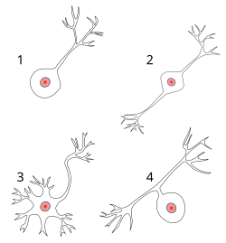
Вспомните, что мы говорили про один длинный аксон и короткие дендриты. Так вот, этот вид нейронов называется мультиполярным, и он — самый «популярный», однако, есть и другие: униполярные (всего один отросток), биполярные (два отростка) и псевдоуниполярные (один отросток, который потом делится на два). Есть и вовсе аполярные(«голые») нейроны. Это предшественники нервных клеток – нейробласты.

Интересно, что униполярные нейроны представлены у человека всего лишь в одном виде: амакриновыми клетками сетчатки глаза. Псевдоуниполярные встречаются гораздо чаще и составляют основную массу спинномозговых чувствительных узлов, о которых мы поговорим чуть позже. Биполярных тоже не так много, и их пул, главным образом, приходится на обонятельные рецепторные клетки. Ну а с мультиполярными и так всё понятно – это универсальные представители нервной системы (например, мотонейроны спинного мозга).
Но, при всей своей важности, строение – это всё же не функции. Каждый нейрон, представляя собой возбуждаемую и возбуждающую клетку (не путать с некими другими физиологическими процессами!), должен своим «настроением» делиться с соседями, иначе сигнал не дойдёт до адресата и не будет обработан и выполнен, что никого, конечно, не устраивает. Поэтому, подобно водителям, въезжающим на платную скоростную трассу, нейроны должны «заплатить», чтобы передать импульс дальше.
Эта «валюта» существует в двух формах: электрической и химической. Второй случай — более частый. А контрольно-пропускные пункты с кассами на автомагистралях воплощаются в синапсах — местах передачи возбуждения с клетки на клетку, то есть местах соединения нейронов. Такие места образуются на специальных выростах на дендритах: дендритных шипиках. Они чаще всего бывают трёх видов: пеньковые, грибовидные и тонкие шипики. Но бывают и другие.

Дендритный шипик — с его шейкой и головкой

Тонкий, грибовидный и пеньковый шипики.
Какие же бывают синапсы?
Реже бывает так. Благодаря ионным каналам в мембране и плотным контактам клеток электрический сигнал без особых усилий перескакивает с нейрона на нейрон и «летит» дальше — пробок нет, оплата принята, водитель доволен. Но это — электрический синапс, или, как еще умничают нейробиологи, эфапс.

Электрические синапсы (эфапсы). а — коннексон (двойная пора) в закрытом состоянии; b — коннексон в открытом состоянии; с — коннексон, встроенный в мембрану; d — мономер коннексина (белка, из которого сделаны коннексоны), е — плазматическая мембрана; f — межклеточное пространство; g — промежуток в 2-4 нанометра в электрическом синапсе; h — гидрофильный канал коннексона.
Но намного чаще случаются ситуации, когда синапс имеет достаточно широкую щель – порядка десятков микрон. То есть перед водителем река, а переправляться придётся на пароме. Здесь вступает в силу химическая «валюта» в виде нейромедиатора, который накапливается в везикулах (пузырьках) пресинаптической мембраны, затем вырабатывается в эквивалентоном силе пришедшего импульса количестве, «переплывает» щель и принимается рецепторами на другом берегу – постсинаптической мебране.
Вот он, универсальный язык нервной системы, а нейроны по типу нейромедиаторов делятся на холинергические, адренергические, ГАМК-ергические и некоторые другие (об этом читайте в следующих выпусках). Исходя из этого, действие, в зависимости от типа нейромедиатора, бывает либо возбуждающим, либо тормозным.

Химический синапс.
Но и это ещё не всё! Есть нейроны чувствительные, которые воспринимают сигнал из внешней или внутренней среды, затем следующие за ними в центральную нервную систему — вставочные, которые обеспечивают ассоциацию в нейронных сетях и могут быть в единичном или множественном числе, и двигательные, которые завершают сигнал действием и иннервируют сократительные или секреторные элементы. Также их ещё можно назвать афферентными (восходящими, двигающимися к центру), интернейронами и эфферентыми (нисходящими, двигающимися к периферии).
«Серый кардинал» нервной системы
Мы поговорили о нейронах, но нельзя забывать и о другой, не менее важной части нервной системы – нейроглии, тем более, что она составляет половину объёма головного мозга и принимает чуть ли не основное участие (как выяснилось в последние годы) в регуляции синаптической передачи, усиливая либо ослабляя сигнал.
Так вот, вся глия по строению, функциям и расположению делится на эпендимную(выстилающую внутреннее пространство цереброспинального канала и желудочков мозга), макро— и микроглию.
Макроглия, в свою очередь, имеет в своём распоряжении целый веер различных подтипов и для центральной, и для периферической нервной системы. Так, в головном мозге она представлена астроцитами, название которых говорит само за себя (большие звёздчатые клетки с большим количеством отростков, которые оплетают нейроны и сосуды), а также олигодендроцитами, которые обеспечивают внутримозговые волокна миелином (по сути, наматываются отростками на аксон — мы уже упомянули о них), многократно увеличивающим скорость передачи импульса.
Периферическая нервная система в основном обходится лишь шванновскими клетками, которые также миелинизируют волокна, но уже за пределами центра, и расходятся по всему организму. И ещё сюда добавляются так называемые мантийные глиоциты или сателлиты, которые образуют оболочку (мантию) вокруг тел нейронов в ганглиях (узлах). Микроглия представляет из себя собственную фагоцитарную систему головного мозга и активируется в основном тогда, когда в нём появляются патологические процессы.

Астроцит.
Но нужно всё-таки подчеркнуть важность глии. Работы по её изучению ведутся не так много лет – буквально два последних десятилетия. Появилась такая рабочая гипотеза (автор — Филип Хейдон [Philip G. Haydon]), согласно которой астроциты, обмениваясь сигналами, активируют нейроны, чьи аксоны находятся от них не только на близком расстоянии, но и сравнительно далеко. Эта активация в итоге способствует высвобождению нейромедиаторов. Таким образом, астроциты регулируют готовность даже отдалённых синапсов к изменению своей эффективности, что представляет собой клеточную основу процессов памяти и обучения.
Сотрудники из лаборатории Бена Барреса (Ben A. Barres, Стэнфордский университет) пошли дальше и открыли специфический белок тромбоспондин астроцитарного происхождения, который стимулирует образование синапсов. Сравнение же головного мозга показывает, что чем более высокое положение занимают животные на «эволюционной лестнице», тем больше в их мозге глиальных клеток по отношению к нервным. Так вот, возможно, что увеличение связности астроцитов может даже повышать способность животных к обучению. Однако это ещё только предстоит доказать.
На острие чувств
В завершение нашего небольшого путешествия внутрь нервной системы разберёмся в том, откуда берутся наши ощущения. Оказывается, здесь строение нервного окончания также имеет самое непосредственное отношение к процессу. Нервные окончания могут располагаться в тканях свободно, могут оканчиваться специальными сенсорными рецепторами, а могут «заключаться» в соединительнотканную капсулу.
Тактильные «граждане» располагаются в слоях соединительной ткани внутренних органов и кожи. Большинство из них – механорецепторы (тактильные, пластинчатые тельца), которые реагируют на какие-либо механические воздействия.
Например, тельца Руффини реагируют на растяжение кожи, тельца Пачини – на давление. Некоторые окончания в эпидермисе «заточены» под регистрацию изменений температуры (тепло – тельца Руффини, холод – колбы Краузе). Есть даже такие рецепторы, которые могут определять изменения рН, рО2 и рСО2.

Поперечное сечение телец Руффини.
Для суставов и мышц есть свои детекторы чувств. К ним относятся мышечные веретёна, сухожильные органы и чувствительные нервные окончания в капсуле суставов.
Источник: портал «Нейроновости»